
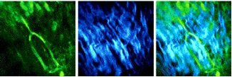
Spectrally resolved SHG-TPEF images of murine footpad bone expressing green fluorescent protein were taken with ultrashort transform-limited laser pulses of 15 fs duration at the objective focus, after compensation via MIIPS. The SHG and TPEF 512 x 512-pixel images were acquired simultaneously in 60 seconds via 16-channel time-correlated single-photon counter. The average laser power on the sample was approximately 10 mW. Images are 150μm x 150μm. BioOptics World 2(2), 23-24 (2009)
Coherent Ultrafast Imaging
Biomedical Imaging:
The fundamental goal of biomedical imaging is the retrieval of structural and dynamical information from the sample, with the greatest contrast, spatial and temporal resolution possible. The workhorse of biomedical imaging is an optical microscope, which readily provides the resolution sufficient to observe sub-cellular organelles. The introduction of lasers and confocal optics brought the resolution scale down to diffraction-limited, i.e. on the order of the laser wavelength. Contrast enhancement comes primarily from the use of fluorescent labeling compounds. Because of their inherent three-dimensional (3-D) sectioning capabilities, nonlinear optical microscopy methods such as two-photon excitation fluorescence (TPEF) and second-harmonic generation (SHG) are gaining popularity. With these methods, the signal is generated within a narrow region near the focus of the microscope objective, and it is well separated spectrally from the laser's excitation wavelength. The laser parameter that makes these nonlinear methods possible is peak intensity, defined as energy per pulse divided by the area at the focus and the laser pulse duration. While the average laser power for nonlinear microscopy imaging is typically 1 to 10 mW, the peak intensity at the focus is as high as 1011 to 1012 W/cm2. These extremely intense fields cause molecules to absorb two or more photons at a time or induce a nonlinear polarization at the sample that gives rise to the second harmonic of the incident frequency.
Publications: 173, 171, 159, 153, 152, 139, 135, 133, 125, 124, 123, 122, 117, 108, 97, 92, 79, 69
Atmospheric Pressure - Imaging Mass Spectrometry:
This is a relatively new and highly promising method being developed in our lab for chemically imaging various kinds of chemical and biological samples at atmospheric pressure. This method offers significant advantages over other imaging techniques by eliminating the need for a laser-absorbing matrix, being suitable for atmospheric pressure sampling, and providing cellular resolution.
Publication: 142